First AId

First AId

Health

First AId

USC physicians are creating byte-size miracles through AI innovation.

December 04, 2024

By Candace Pearson

A custom heart or vascular stent for everyone who needs one. Your own stem cells providing the material for 3D-printed organs. Spray-on skin. A powerful diagnostic partner that enables more equitable health care. This is the promise of artificial intelligence (AI), now a full member of any health care team — and USC is leading the way.

AI excels at analyzing large amounts of datasets, and it has applications across every aspect of health care. At USC, researchers and clinicians are using AI to minimize risks and improve patient outcomes, create precision surgical techniques, hasten drug discovery and interpret medical images with impressive accuracy — all treatments thought out of reach just a few years ago.

“In 1980, medical knowledge doubled every seven years,” says Summer Decker, founding director of USC’s Center for Innovation in Medical Visualization. “Today, it more than doubles every 72 days. AI enables us to track patterns we couldn’t see before, because it would be almost impossible to analyze that volume of data. It will help us start seeing those patterns earlier so we can begin addressing problems earlier.”

Already, AI-powered wearables monitor patients’ health metrics 24/7, while 3D printing produces replicas of human organs for study in various applications such as transplantation, disease modeling and drug testing. “This isn’t science fiction,” says Inderbir Gill, executive director of Keck Medicine’s USC Urology, who himself regularly performs AI-assisted robotic surgery. “How can you not be impressed by the potential of AI? It’s coming at us at breakneck speed.” Gill is also Distinguished Professor and chair of the Catherine and Joseph Aresty Department of Urology at the Keck School of Medicine of USC, which hosted the first AI West Med Symposium in February.

AI also helps solve a more mundane — and yet very real — health care challenge: physician burnout from administrative tasks. By using AI to update electronic health records, clinicians can free up time for patient care.

“The opportunity of AI is to take this massive amount of data and improve humanity,” says Steve Shapiro, USC’s senior vice president of health affairs. “By having all this incredible wealth of data and AI algorithms, we’re going to be able to ignite discovery.

“Once we have all this information, we can generate hypotheses, take that to the lab and make real discoveries,” Shapiro adds. “Our scientists, students, clinicians can ask the most important questions and challenge dogma.”

A strong partnership among Keck Medicine of USC, the university’s health schools and the USC Viterbi School of Engineering, alongside national and global colleagues, provides the ingredients for medicine that ultimately may help millions worldwide.

But will AI replace our doctors? Not at all, says Carolyn Meltzer, dean of the Keck School of Medicine. For all of AI’s capabilities, algorithms can’t do everything: Compassion, ethics and creative, nonlinear thinking remain strictly human. “It’s not that AI will replace the physician,” Meltzer says. “It’s that the physician who uses AI will replace the physician who does not use AI.”

Meet some of the physicians at USC who are deploying AI tools to transform healing.

From the Virtual to the Physical
asdaskldjlkasjd
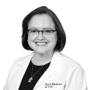
Summer Decker

Summer Decker, founding director of USC’s Center for Innovation in Medical Visualization and professor of Clinical Radiology, Surgery and Pathology, creates 3D-printed models of human anatomy — for surgical planning, teaching, medical device development and patient care. Leveraging AI, she says, can improve print quality, detect and correct errors before they happen and make 3D printing more accessible by simplifying the process.

“With 3D printing, we can reduce the time needed for surgery, minimize the risk to patients and know exactly what size surgical device or replacement part to use for each individual. It gives surgeons a road map inside the body with real-time 3D views of a patient’s anatomy. There’s so much we can do with 3D information, whether you’re a surgeon planning an operation or a student learning a procedure. Our patients are the ones who will benefit. When I looked at where I wanted to be to push this technology, the answer was USC because it’s a world leader.

“Our lab holds several patents on a technology that will seed a 3D print with demineralized bone matrix, which can form a custom internal scaffold for facial injuries in kids. Unlike a metal plate, the print and tissue will grow with them. In bioprinting, which is already here, you take a person’s stem cells and reprint their anatomy. Of course, certain organs are easier than others to print. The brain is a long way away — if ever. AI will give us the tools to be better at what we do. We’ve just got to drive it.”

Building Custom Stents
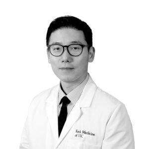
Sukgu Han

Sukgu Han is a vascular surgeon and biomedical engineer who designs and crafts custom stents for his patients. He is a professor of clinical and neurological surgery and chief of the Division of Vascular Surgery and Endovascular Therapy at Keck Medicine. He also serves as the co-director of the Comprehensive Aortic Center at Keck Hospital and program director of vascular surgery residency and fellowship at Keck School of Medicine.

“Everybody’s anatomy is a little different. An aneurysm — a bulging, weakened area in the aorta — can distort the anatomy as the blood vessel twists on itself. Having the ability to tailor each patient’s stent to fit their anatomy and protect the aorta can make surgery safer. I began making complex, custom aortic stent grafts for my patients 10 years ago. I’ve now done more than 500 — a lot of it I’ve done manually. The next phase will be automated AI analysis, which will speed the process of sizing the stents in three dimensions to fit each patient.

“We recently launched a project using an AI vessel-segmentation algorithm that will compare a patient’s CT scan with their previous scan to determine what is happening in their aorta in much greater detail than how we’re doing this currently — which is basically a radiologist or surgeon putting the images side by side and hoping to detect any changes that make a difference in the patient’s care, but can easily be missed due to human error.

“We haven’t even scratched the surface in tailoring each patient’s treatment to a deeper granularity. No one has the technology to 3D print some of these custom stents — yet. That may be coming our way sooner than expected. Next is printing cardiac structures like heart valves for implantation. Who knows what we’ll be talking about in five years?”

Preventing Avoidable Injuries
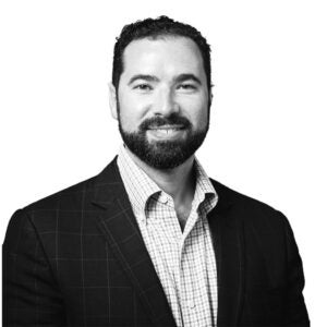
William Padula

William Padula is an advocate for using AI to better predict pressure injuries, commonly known as bedsores. He is assistant professor of pharmaceutical and health economics at the USC Alfred E. Mann School of Pharmacy and Pharmaceutical Sciences.

“More people die from pressure injuries acquired in the hospital than from car accidents. The annual cost of hospital-acquired pressure injuries in the United States is $30 billion and 60,000 deaths. By my calculation, we could probably prevent most injuries in a smart hospital infrastructure using data science for $10 billion to $15 billion and eliminate $30 billion in waste and save many lives. This would give us a lot of financial bandwidth to do other things, such as gene therapy for sickle cell disease or for kids with cystic fibrosis.

“In the 1980s, nurses began using a paper-based tool to document pressure injury risk factors on a scoring sheet — an antiquated method by today’s capabilities. USC is collaborating with Johns Hopkins University and UH Cleveland Medical Center; we developed a risk-assessment model using machine learning, which increased the accuracy of risk prediction to 74% — a more than 20% jump over human methods.

“An unbiased machine-learning tool based on data consistent across all patients — whether sensory data or blood tests — will enable us to better predict the risk each patient faces of developing a pressure injury or other adverse health outcomes. Through AI, we can make health care more equitable and more focused on prevention and treatment. That’s the potential of AI.”

Offering ‘An Extra Set of Eyes’
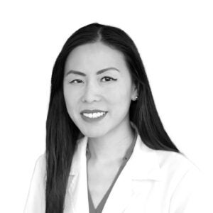
Melinda Chang

Melinda Chang, assistant professor of clinical ophthalmology at USC Roski Eye Institute, part of Keck Medicine of USC, wants to improve diagnosis of swollen optic nerves in kids, a potentially serious neurologic problem. She believes AI can reduce the need for diagnostic procedures that require sedation of children.

“Swollen optic nerves, known as papilledema, can be a sign of a brain tumor, hydrocephalus or meningitis. It’s one of the most common reasons kids are referred to our pediatric neuro-ophthalmology clinic. Complicating the diagnosis is pseudopapilledema, in which the optic nerves also appear swollen but are essentially benign.

“No single imaging technique can accurately differentiate between the two conditions on its own. Even combining them all might not be accurate enough. A full workup requires an invasive lumbar puncture and sedation in kids, which we want to avoid. We recently initiated a multicenter study comparing human diagnoses and an AI model programmed to scrutinize minute details in photos of the back of the eye.

“The human experts did very well in distinguishing severe papilledema from pseudopapilledema, but AI did much better. In cases of mild papilledema, which is harder to differentiate, AI achieved an accuracy of approximately 70% and sensitivity close to 90%, far surpassing the performance of human experts, whose accuracy ranged from 53% to 59% and sensitivity from 39% to 53%. For us, that means AI has promise in these difficult cases in potentially reducing the rate of kids undergoing unnecessary tests and missed neurological problems. AI won’t replace doctors but will serve as an extra set of eyes.”

Cancer Care at Home
Jorge Nieva

Jorge Nieva is exploring at-home care for patients with non-small cell lung cancer, which is made possible by telemedicine and remote monitoring. A Keck Medicine of USC medical oncologist and lung cancer expert, he is associate professor of clinical medicine, Keck School of Medicine of USC.

“These days, 30% to 40% of my lung cancer patients can be treated with a pill. Some cancers, like non-small cell lung cancer, still require injections. Our new clinical trial deploys nurses to patients’ homes to administer immunotherapy medication under the skin. During the pandemic, the use of telemedicine exploded, and patients appreciated the convenience, especially in Los Angeles, which can be hard and stressful to navigate. The trial also relies on telemedicine visits and remote monitoring. It’s like a modern house call. My excitement about digital tools like these is having access to real-time and self-reported data on the patient’s vital signs, movement data and symptom management. It leads to better decision-making.

“Some applications of AI in health care will be very important, such as assistance with the analysis of medical images and pathology slides, and chatbots for triaging patient communications. When seeking to improve the health of a population through better care coordination and patient engagement, AI can help ensure patients get timely cancer screenings and follow-up. But we’re not at the day yet when someone will type their symptoms into a box, receive a diagnosis and a prescription in the mail.

“With something as critical as a diagnosis, you’re always going to want a human who has thought about the problem enough that they’re willing to say, ‘No, I’m responsible. The buck stops with me.’”

From Skin-Like Wearables to Gut Clues
Yasser Khan

Yasser Khan invents AI-powered wearable, implantable and ingestible medical devices. He is an assistant professor of electrical and computer engineering at the USC Viterbi School of Engineering and the USC Institute for Technology and Medical Systems Innovation, a joint Keck School of Medicine of USC-USC Viterbi initiative.

“Imagine using AI-enabled devices to track your physical and mental health. By printing electronics on plastic and rubber, wearables become so thin, flexible and stretchable, they behave like a second skin. One study I was involved in developed a ‘bandage’ that can measure tumor growth within the width of one human hair. Now we’re designing a wearable sensor in patch form with physiological and bio-chemical sensing capability to help classify mental states. And the skin-like electronics revolution is just starting.

“In addition, my lab is focused on implantables and ingestibles — the latter is brand-new work out of USC. We are now designing a combination wearable/ingestible system that could someday serve as a ‘Fitbit for the gut’ for early disease detection. It combines a smart pill that can measure gases, chemical markers and neurotransmitters, plus a wearable system (essentially a coil over your shirt) with high resolution to follow the capsule. There’s no way now to measure these things noninvasively. The hope is this capsule someday can carry a chemotherapy drug and be released in the exact location to target the cancer.”

Hope for Diabetics
David G. Armstrong

David G. Armstrong is a professor of surgery and neurological surgery at the Keck School of Medicine of USC, a podiatric surgeon and limb preservation specialist with Keck Medicine of USC. A diabetic wound care expert, he works within a coordinated program to treat diabetic foot ulcers and other chronic wounds. Their work is merged with technologies such as injectable sensors, gene-therapy- directed wound healing and Bluetooth-enabled artificial blood vessels.

“Every second someone develops a diabetic foot ulcer. Half of these people get a foot infection, and 20% end up in the hospital. Every 20 seconds there’s an amputation. We believe virtually all of this is preventable. That’s why we developed the predictive Smart Boot, a wearable technology supported by AI-based algorithms that can help diabetic patients recover from dangerous foot wounds. It can identify not only whether people are wearing it but how fast they move, the steps they take, if they’re unsteady or about to fall.

“Right now, there’s a fundamental blurring of the lines between consumer electronics and medical devices. With our Caltech colleagues, we’re bringing smart bandages that sense and respond to inflammation or infection to clinical use. Having worked with augmented/virtual reality for two decades, I’m also excited about the Apple Vision Pro opening up possibilities for surgical procedures, medical training and care.

“These devices capture the imagination — and they’re getting better at helping us look after our patients better and drive medicine forward. By marrying our humanity with the technology, we can effect positive change on ourselves and the planet. We don’t have to predict the future. It’s all happening now.”

Cell by cell: Rebuilding the body

Cell by cell: Rebuilding the body

Health

Cell by cell: Rebuilding the body

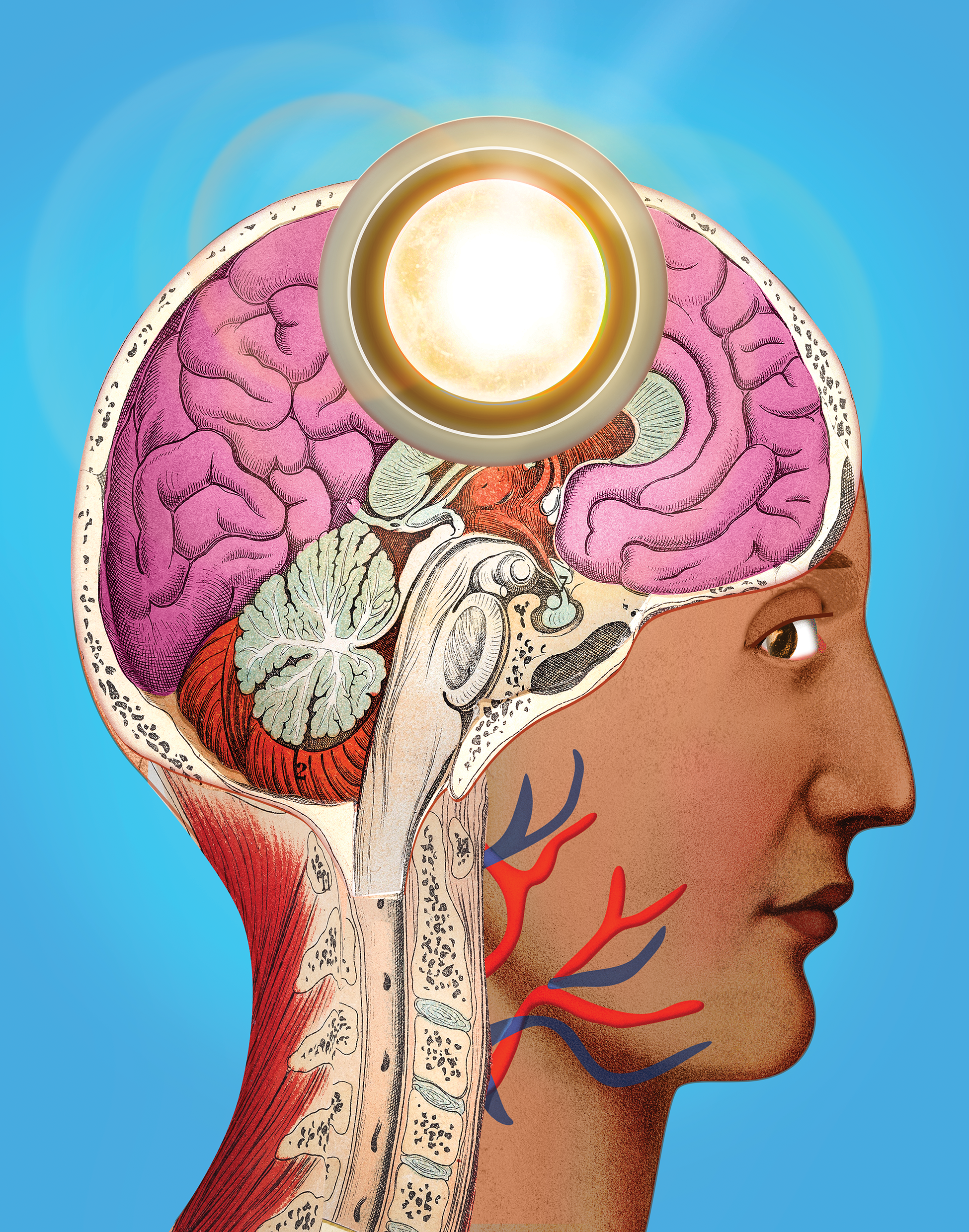
USC researchers are revolutionizing how we treat disease by harnessing stem cells as “living medicine.”

December 04, 2024

By Rachel B. Levin

Anyone who’s healed from a cut or a scrape has witnessed the incredible regenerative power of stem cells. These cells can create identical copies of themselves, creating new cells and tissues that replace damaged ones.

Stem cells are active in some areas of our body throughout our lives, like the skin and blood. But in many critical organs, including the heart and kidneys, stem cells are absent. When such tissues are damaged due to aging, injury or disease, they don’t regenerate, leading to devastating health consequences.

USC researchers are at the forefront of an emerging field called “clinical regenerative medicine,” which taps stem cells’ restorative powers to tackle some of the hardest-to-treat diseases, ranging from heart failure to blindness.

“We now have the ability through stem cells to generate replacement cells that we can use as therapeutics to rebuild the human body,” says Charles (Chuck) Murry, a renowned expert in regenerative heart medicine. In August, Murry joined Keck School of Medicine of USC as the new head of USC Stem Cell, chair of the department of stem cell biology and regenerative medicine, and director of The Eli and Edythe Broad Center for Regenerative Medicine and Stem Cell Research.

Launched in 2013, USC Stem Cell is a universitywide initiative that connects over 100 research and clinical faculty in multiple disciplines across USC and Children’s Hospital Los Angeles (CHLA) with the common goal of translating basic stem cell science into clinical therapies. It has matured over the past decade with support from the Eli and Edythe Broad Foundation as well as the California Institute for Regenerative Medicine (CIRM), a state organization created to accelerate stem cell research.

USC Stem Cell collaborators are employing stem cells to grow organ and tissue replacements, halt or reverse the progression of life-threatening diseases and create living models of human organs in the lab, providing novel platforms to screen for disease-fighting drugs.

“Clinical regenerative medicine is going to have an impact on par with antibiotics or vaccinations,” Murry says. “It’s going to be revolutionary.”

The stem cell projects currently underway across USC will transform treatments from our skull to our knee and many organs in between. As with the classic board game Operation, we’ve broken down a selection of these projects by body part — funny bone not included.

SKULL

The bones of our skull protect one of the most important human organs: the brain. When those bones are compromised either because of a congenital condition, injury or surgery, the defect may affect not only the appearance of the skull but also brain function itself. Since skull bones do not regenerate after infancy, surgery is often needed to repair defects.

Yang Chai, University Professor, interim dean and associate dean of research at the Herman Ostrow School of Dentistry of USC, treats craniofacial birth defects. His work inspired him to develop an innovative treatment option for patients with significant defects — essentially holes — in the calvarial bones at the top of the skull.

Typically, these holes are surgically closed with a metal or plastic plate. For pediatric patients, neither option is ideal because a synthetic plate will not grow in concert with the child’s developing brain. Many of these implants fail within 20 years.

Collaborating with Yong Chen, professor of aerospace and mechanical engineering and industrial and systems engineering at the USC Viterbi School of Engineering, Chai designed a “living” implant for skull bone regeneration. It’s a 3D-printed scaffold made of a mineral found in teeth and bones called hydroxyapatite. The scaffold can be customized to fit a patient’s defect precisely and seeded with a patient’s own bone marrow-derived stem cells.

In animal models, as the stem cells mature, they grow and integrate with the surrounding skull to cover the defect within about six months. The new bone becomes as strong as the native bone. Chai and his collaborators — including Mark Urata, professor of clinical surgery at the Keck School of Medicine of USC — are poised to begin clinical trials in humans.

“Ours is going to be a biological solution for a biological problem, instead of a mechanical solution [i.e., a synthetic plate] for a biological problem,” Chai says.

BRAIN

Studying human brain development is important in understanding how neurodevelopmental issues such as autism spectrum disorder arise and progress. But traditional research methods have proved insufficient for observing the human brain as it develops. Current brain imaging techniques don’t offer a resolution that is high enough to understand brain functionality at the cellular level. Postmortem human brains don’t provide a window into early growth, and animal models don’t possess the complexity of the human brain.
“The best way to access human brain development is really to take advantage of human stem cells,” says Giorgia Quadrato, assistant professor of stem cell biology and regenerative medicine at the Keck School of Medicine of USC.

Quadrato has pioneered a way to grow masses of brain cells in the lab from stem cells. Within six months, these cells mature into tiny structures (“brain organoids”) about two millimeters in diameter that contain all the various cell types of specific brain regions. The organoids allow Quadrato’s team to study the cells’ development in real time, model neurodevelopmental diseases and screen for potential new treatments.

One of her lab’s most noteworthy innovations is an organoid model of the cerebellum, a brain region that plays a role in movement, cognition and emotion. In certain neurodevelopmental and neurodegenerative disorders like autism spectrum disorder and cerebellar ataxia, cells in the cerebellum called Purkinje neurons degenerate. Quadrato’s team is the first to succeed in growing Purkinje cells with all the features of functional neurons in a human system.

While the cerebellum models were grown from healthy cells, Quadrato has also grown organoid models of the cortex — a brain region involved in cognitive function — derived from the cells of a patient with a disease-causing variant of the SYNGAP1 gene. This variant is one of the top risk factors for autism spectrum disorder. The lab’s organoid models shed new light on how the variant disrupts early development of the cortex.


EYES

As we age, changes to the retina, the delicate tissue at the back of our eyes, can lead to vision loss. Photoreceptor cells that sense light and send signals to the brain can deteriorate due to inflammation, genetics and lifestyle habits such as smoking. At the center of the retina, the macula, which controls straight-ahead vision, is particularly vulnerable to damage.

Age-related macular degeneration (AMD) is the leading cause of severe vision loss among people over 50 in Western countries. Because there’s no known way to regenerate photoreceptors, AMD has had no effective treatments — until now.

Mark Humayun, University Professor of ophthalmology at the Keck School of Medicine of USC and co-director of the USC Roski Eye Institute, and his collaborators are using stem cells as a gateway to restoring vision in people with dry AMD, the most common form.

The disease affects not only photoreceptor cells but also a vital group of cells that protect them from damage called retinal pigment epithelium (RPE) cells. Humayun and his team developed a tiny patch filled with RPE cells derived from stem cells that can be surgically implanted in the eye, where the young cells can shield patients’ photoreceptors from further degeneration.

Clinical trials of the patch are run by Regenerative Patch Technologies, the company he co-founded with Dennis Clegg and the late David Hinton. In the first phase of the trial, 27% of patients experienced improved vision.“We found that these young RPE cells are very robust,” Humayun says. “They survive much better than adult-aged RPE. Patients who improved are still keeping the same level of vision three years later.”

This fall, during the trial’s second phase, the patch will be tested in a larger number of dry AMD patients. Among them will be patients with less severe disease than patients in the first phase. Humayun expects that in this phase the patch will improve vision in even more patients and to a greater extent.

MOTOR NEURONS

Amyotrophic lateral sclerosis (ALS) — often called Lou Gehrig’s disease — is a devastating neurodegenerative disease with no known cure. It affects motor neurons, which are nerve cells in the brain and spinal cord that control voluntary muscle movement and breathing. Over time, as motor neurons degenerate, the brain can no longer command muscle movements, and people with ALS may lose their ability to speak, eat, walk and breathe.

Finding universally effective treatments has been difficult because the disease’s root causes are not fully understood. While having a family member with ALS increases your risk of developing it, most cases occur with no family history or clearly associated risk factors. “In these cases, we believe that multiple gene mutations come together in one person,” says Justin Ichida, an associate professor of stem cell biology and regenerative medicine at the Keck School of Medicine of USC whose research is centered on fighting the disease.

Stem-cell technology has provided Ichida an avenue for new leaps in discovery. He and his team reprogram adult skin or blood cells from an ALS patient into stem cells. These cells are then coaxed into developing into motor neurons that are genetically identical to the patient’s own motor neurons. The process has allowed Ichida’s lab to test thousands of compounds to see how the neurons respond.

One of Ichida’s most exciting discoveries is that inhibiting a protein called the PIKFYVE kinase improves the survival of motor neurons derived from patients with varying types of ALS. His startup company, AcuraStem, developed a druglike molecule to suppress this kinase and licensed it to a pharmaceutical company, which is now developing it into a drug for clinical trials. “This could be applicable for essentially all ALS patients, rather than just a subset that have a certain type of mutation,” Ichida says.

EARS

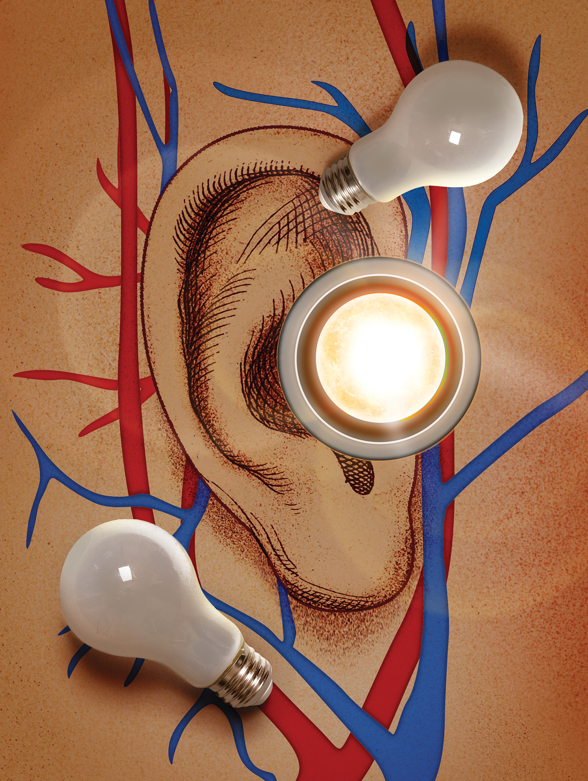
Inside your cochlea — the spiral structure inside your inner ear — a few thousand tiny sensory receptors with bundles of hairlike protrusions (so-called “hair cells”) perform a function essential for hearing. They convert the mechanical vibration evoked by sound waves into electrical signals that your brain interprets. When they’re damaged either from wear and tear or disease, hair cells can’t be normally replenished.
That’s true in humans, mice and other mammals. But in non-mammalian vertebrates, such as songbirds, damaged hair cells regenerate in a matter of weeks. The difference comes down to the activity of stem cells called supporting cells.

“The first thing a supporting cell needs to do is sense that there is an injury and divide into progeny that will mature into hair cells,” says Ksenia Gnedeva, assistant professor of otolaryngology – head and neck surgery and stem cell biology and regenerative medicine at the Keck School of Medicine of USC. “That doesn’t happen in mammals. The very first step is blocked.”

Gnedeva discovered that a molecular cascade called the Hippo signaling pathway acts like a brake on that first step in regeneration in mammals. Then, she identified a small molecule that blocks the main enzyme in the pathway called the LATS kinase that can switch the cascade from a brake to an accelerator.

“When we applied the kinase inhibitor to inner ear sensory organs from mice, we saw something that had never been published before,” Gnedeva says. “All of the supporting cells were now able to reenter the cell cycle,” meaning that they began actively dividing in the petri dish. Remarkably, some of them grew into hair cells.

Gnedeva’s lab is now investigating how to effectively deliver this compound into the inner ear to restore hair cells in living mice. The hope is that the therapy may one day work to regenerate hair cells in humans and restore hearing to those with severe hearing loss.


KNEES

In your joints, a strong, flexible connective tissue called cartilage provides padding between bones and acts as a shock absorber and lubricant. Osteoarthritis occurs when the cartilage is compromised due to injury or aging, causing the bones to rub against each other, often painfully.

Very few stem cells are present in adult cartilage, meaning the tissue doesn’t naturally regenerate. Historically, treatments for osteoarthritis have been limited to painkillers and invasive surgery to replace damaged joints. But Denis Evseenko — professor of orthopedic surgery, stem cell biology and regenerative medicine, vice chair for research of orthopedic surgery and director of skeletal regeneration at the Keck School of Medicine of USC — and his collaborators have innovated new stem-cell-based treatments that promise to make osteoarthritis pain a thing of the past.

One is a “regenerative pouch” to replace cartilage that’s been damaged by a fall, sports injury or other trauma. The pouch, which is being developed by Evseenko’s startup company Plurocart, contains hundreds of thousands of young cartilage cells derived from stem cells. “It’s a little reparative structure that you can surgically deliver right into the cartilage defect,” Evseenko says.

In animal models, the pouches have shown phenomenal results: The juvenile cells grow into mature cartilage, preventing osteoarthritis. Evseenko is now planning the first clinical trial in humans with knee injuries.

Evseenko’s lab has also created a therapy for age-related osteoarthritis that is being developed by his startup company CarthroniX. As we age and cartilage is progressively lost, painful inflammation can set in. “Chronic inflammation is a signal for local stem cells that it’s a bad environment and they should not build new tissue,” Evseenko says.

He identified a compound that disrupts a key immune receptor’s over-activation due to inflammation. When this compound is injected into the knees of patients with mild to moderate osteoarthritis, inflammation is suppressed, stem cells are activated to slowly regrow tissue and pain is significantly reduced.

HEART

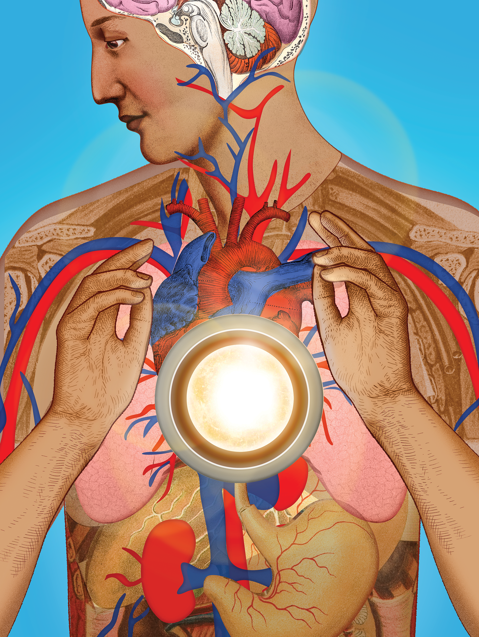
After a heart attack, many people go on to lead productive lives — but the heart muscle itself is forever changed. Since adult heart cells do not regenerate, the scar tissue formed in areas of the heart attack can compromise the heart’s ability to pump. Over time, reduced function can lead to heart failure, depending on the amount of scar tissue and the size of the heart attack.
Murry, the head of USC Stem Cell, has discovered a way to use heart cells derived from stem cells to strengthen hearts damaged by scar tissue. “By transplanting these heart muscle cells, we can rebuild the heart and restore its contractile function,” Murry says.

He likens the process to growing a garden: The injured heart becomes the soil where stem-cell-derived heart cells are planted. The “roots” are the connective tissue and blood vessels that the new cells call in to feed them. Murry anticipates that clinical trials of the transplanted cells will begin within the next couple of years.

Megan McCain, associate professor of biomedical engineering at the USC Viterbi School of Engineering and of stem cell biology and regenerative medicine at the Keck School of Medicine, has innovated complex living tissue models from human stem cells that allow her to visualize heart scar tissue formation in real time.

Her team seeds quarter-size silicone wafers with stem cell-derived heart muscle cells. As the cells grow, they align and begin to beat together much as they would in an actual human heart. McCain can then induce what she calls a “heart attack on a chip” — by selectively depriving some of the cells of oxygen — and observe how the cells remodel themselves after the injury.

“The next step is to develop interventions to change how the heart heals itself,” McCain says, to reduce how much scar tissue forms in the first place.

Vaughn Starnes, chair and distinguished professor of surgery at the Keck School of Medicine and founding executive director of Keck Medicine’s USC Cardiac and Vascular Institute, is exploring stem cells as a treatment for children born with a congenital condition called hypoplastic left heart syndrome. In those affected, the left side of the heart is underdeveloped, leaving the baby with one, instead of two, functioning ventricles to pump blood. Over time, the single ventricle fails in 10% to 15% of patients.

Early evidence suggests that when stem cells from a baby’s own umbilical cord blood are differentiated into heart cells and injected into that ventricle, they strengthen the muscle and improve its function — potentially reducing the chances of failure down the line.

KIDNEY

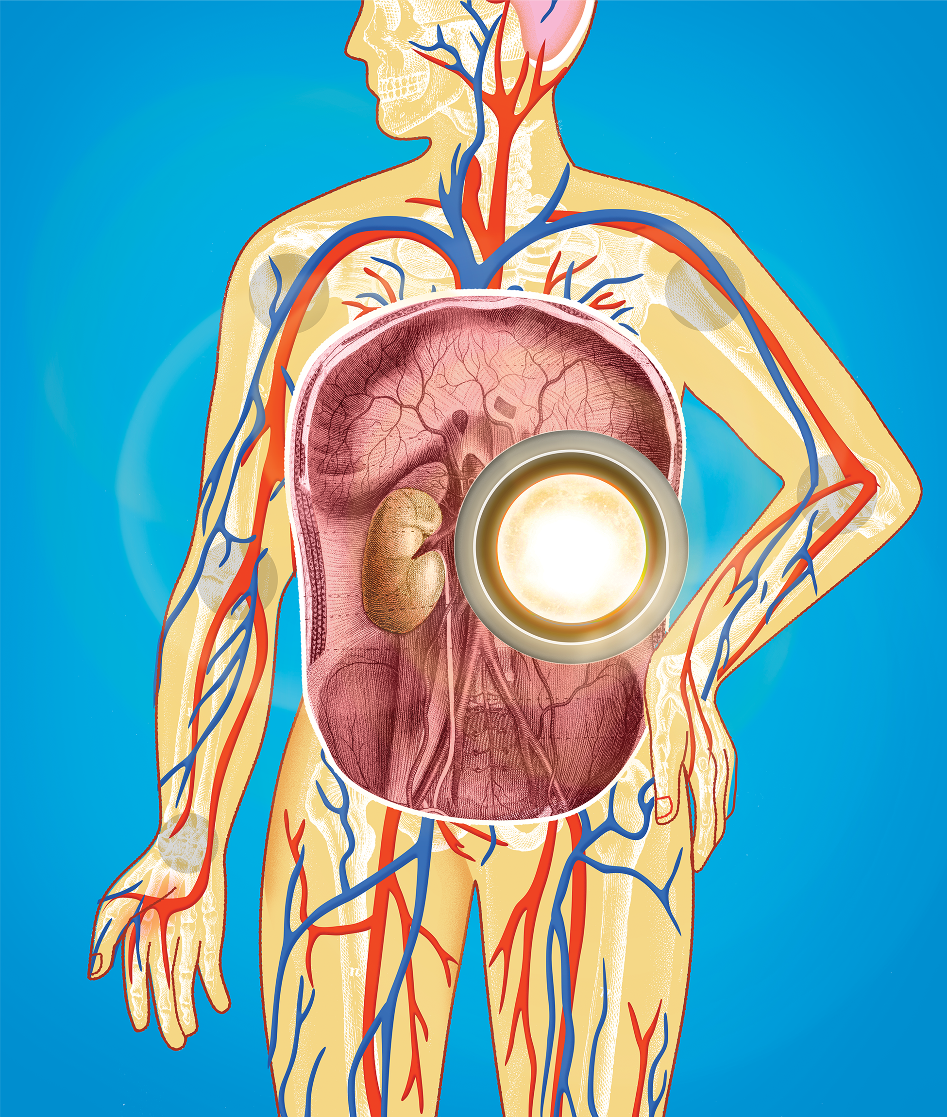
Our kidneys are vital organs to sustain life. They filter waste products from our blood and produce urine we excrete. Because of the increasing prevalence of diseases that injure the kidneys, including hypertension and diabetes, chronic kidney disease is on the rise in the United States, affecting one in seven adults.

Kidney transplants can save lives — but the demand for donor kidneys far exceeds the supply. Each day, an estimated 13 Americans die waiting for a kidney transplant.

Zhongwei Li, assistant professor of medicine and stem cell biology and regenerative medicine at the Keck School of Medicine of USC, has pioneered a “synthetic kidney” using human stem cells that may soon provide an alternative to kidney transplants from a donor.

The architecture of a human kidney is complex. A million functional units called nephrons organize themselves into a treelike structure to drain the urine they produce. Once injured, nephrons can’t regenerate. Li developed a method for turning stem cells into kidney progenitor cells, the cell type necessary for kidney development in a human embryo.

“By using those building-block progenitor cells, we have successfully assembled a treelike structure with nephrons attached to the structure in a petri dish,” Li says.

His team has demonstrated that these “kidney organoids,” when transplanted into animal models, can grow into synthetic kidneys that produce urine just like natural kidneys. Li anticipates that within five to 10 years, the technology will be advanced enough for clinical trials in humans.

As he works with collaborators in fields including kidney development, 3D bioprinting and kidney physiology to perfect the synthetic kidney’s design, Li stresses that the power of his innovation comes from nature itself.

“Progenitor cells are hardwired to build a treelike structure,” Li says. “We direct the cells to do what they’re programmed to do.”

PANCREAS

For the more than 38 million Americans with diabetes, the disease significantly affects daily life and poses long-term health risks. While it can be managed with medication, diet and lifestyle, there’s currently no cure.

Diabetes occurs when your blood sugar is too high, either because your pancreas makes little or no insulin (Type 1 diabetes) or your cells don’t use insulin properly (Type 2 diabetes). In the latter condition, your pancreas may be producing insulin — just not enough to keep your blood sugar in the normal range.

“Whether it’s Type 1 or Type 2 diabetes, fundamentally you need more functional insulin cells,” says Senta Georgia, associate professor of pediatrics and stem cell biology and regenerative medicine at the Keck School of Medicine of USC. “For a number of years, my lab has been working on ways to make insulin cells from stem cells, trying to understand the barriers that exist and improve the processes that underlie how we make these cells.”

Georgia aims to support the development of clinical regenerative therapies for diabetes. These may one day include transplanting stem cell-derived insulin cells into the pancreas, coaxing a patient’s pancreas to make more of its own insulin cells or improving the function of existing insulin cells.

One of her lab’s current areas of focus is investigating why COVID-19 infections sharply increase the risk of developing Type 2 diabetes. Her team is using stem cell models to understand the virus’ impact on insulin cell function and survival, with an eye toward identifying and blocking pathways to injury.

They’re also collaborating with clinicians at CHLA to study the cells of pediatric patients with genetic forms of diabetes. A recent project illuminated the critical importance of a gene called NEUROGENIN3 in enabling stem cells to mature into insulin cells. Children with a mutation in this gene develop a severe form of diabetes.

CELLS

While many USC researchers are employing stem cells to regenerate damaged tissues and organs, Ian Ehrenreich — divisional dean for life sciences and professor of biological sciences at the USC Dornsife College of Letters, Arts and Sciences — is pioneering new ways to modify stem cells themselves.

Genetically modified stem cells hold great promise for therapeutic purposes. Ehrenreich points to one recently developed example: the first FDA-approved gene therapy for patients with sickle cell anemia, a blood disorder caused by a genetic mutation. In this therapy, the blood stem cells of patients are modified by genome editing and transplanted back into patients’ bone marrow, where they produce a protein that prevents the “sickling” of red blood cells.

“The type of genetic engineering involved in this therapy is simple,” Ehrenreich says. “They’re breaking one genetic element. But a lot of things we’re going to need to do [to develop new stem-cell therapeutics] are going to require more sophisticated genetic engineering. That’s the realm where my lab works.”

He and his team have developed new techniques for building large pieces of DNA (synthetic chromosomes), involving less time and cost than ever before possible. “These big DNA constructs will be important for reprogramming stem cells to serve particular tasks,” Ehrenreich says. DNA synthesized using these techniques could also be added to a stem cell to compensate for genes that a patient is missing.

One of Ehrenreich’s intracampus collaborations is with Leonardo Morsut, assistant professor of stem cell biology and regenerative medicine at the Keck School of Medicine of USC. Morsut engineers stem cells to divide in particular ways to grow tissues of desired structure and function. Employing large synthetic chromosomes from Ehrenreich’s lab “might enable more complex programming of these cells, which could be beneficial in eventually making [synthetic] organs,” Ehrenreich says.

Turn back the clock on aging

Illustration by Drue Wagner

Illustration by Drue Wagner

Health

Turn back the clock on aging

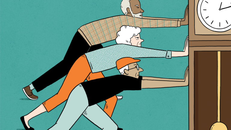
There’s no way to stop the march of time, but innovative research by USC scholars points the way to a longer, healthier and more vibrant life.

December 04, 2024

By Rachel B. Levin

About 10 years ago, when George Salem was in his mid-50s, a friend invited him to play a round of golf. He accepted the invitation — even though the associate professor in the USC Division of Biokinesiology and Physical Therapy had never taken golf seriously. A former college football player, he favored more vigorous sports like snowboarding, basketball and rugby.

But when he joined his friend’s foursome, Salem found himself riveted by the action on the greens and fairways. The course was crowded with silver-haired players, many in their 80s and 90s. He struck up conversations with individuals who had survived cancer and strokes, undergone hip and knee replacements, and were grappling with Parkinson’s disease. And during play, these older golfers left Salem in the dust.

“I was amazed at how much better at golf they were than I was,” he says. “But more importantly, I was amazed at how they could get in and out of a sand trap, climb hills, track a ball and mentally strategize which club to use and how to shoot.”

An expert in exercise and aging, Salem wondered if golf itself was helping to keep these players physically robust and cognitively sharp despite their health challenges. Right there on the links, a new research direction was born.

For millennia, humans have been searching for ways to extend life and remedy the ravages of aging, pursuing mythical springs, miracle elixirs and science-based biohacks. In recent decades, as life expectancies have increased, the desire to thrive as we age has only intensified.

But quality-of-life challenges still loom for an older adult population growing at an unprecedented pace. This includes rising rates of age-related chronic diseases, social isolation and elder abuse.

Now an avid golfer and director of the USC Institute for Therapeutic Golf Science, Salem is among the many USC scholars in wide-ranging fields whose innovative research is meeting these challenges head-on — and redefining the path to longevity in the process. Whether leveraging science and technology or harnessing the power of community, USC researchers are making leading-edge discoveries that can guide the choices we make today for a healthier and happier tomorrow.

“USC is uniquely positioned to chart new territory in our understanding of longevity and healthy aging,” says Pinchas Cohen, dean of the USC Leonard Davis School of Gerontology, which is the nation’s first and largest educational institute on aging. “We are unmatched in our constellation of resources to support patients, families and caregivers and develop visionary new ways to increase both lifespan and healthspan — years spent free of disease.”

You Are What You (Don’t) Eat
Illustration by Drue Wagner

You Are What You (Don’t) Eat

THOUGH THE PASSAGE of time is a constant, the rate at which we age varies from one person to another.

Scientists make a distinction between your chronological age and your biological age, the latter being a measure of the functional state of your body’s systems and damage that’s occurred to your cells and tissues. If your biological age is greater than your chronological age, you’re more likely to develop age-related diseases including dementia, stroke and more. While genetics influence the pace of biological aging, making healthy lifestyle choices can slow it down — or possibly put it in reverse.

One of the most important choices is what you put on your plate. Exploring a century of research on long-lived populations around the world, Valter Longo, professor of gerontology and biological sciences at USC Leonard Davis, developed recommendations for a “longevity diet” that is rich in legumes, whole grains, vegetables, nuts, olive oil and some fish, and low in red and processed meats, sugar and refined grains.

But his research also demonstrates the benefits of taking breaks from eating abundantly. A recent study Longo conducted with Sebastian Brandhorst, associate professor of gerontology, and other collaborators reveals that periodically adopting a “fasting-mimicking diet” (FMD) — which contains ingredients that simulate fasting and drastically limits calorie intake for five days — can shave years off your biological age. In the study, adults who completed three FMD cycles over several months slashed their biological age by an average of 2 1/2 years, and their insulin resistance and other pre-diabetic markers improved.

“Humans and many other organisms evolved during long periods when we didn’t have food,” Longo says. His lab’s animal studies suggest that when our hunter-gatherer ancestors feasted after a period of famine, a process called cellular reprogramming kicked in so that shrunken cells, organs and tissues could be rebuilt and restored. “The fasting-mimicking diet is tapping into this ability of the system to detect damage and then begin a process of self-repair and regeneration,” he says.

Longo has shown that in mice, periodic FMD cycles reduce harmful inflammation, kill cancer cells and improve markers of Alzheimer’s disease in the brain. Early research suggests they may do the same in people. Longo’s discoveries have been leveraged by his company L-Nutra to create FMD kits for general health and tailored to specific diseases including diabetes, cancer and Alzheimer’s.

Illustration by Drue Wagner

Teeing Up for Play Time

OUR PREHISTORIC predecessors’ survival depended upon physical fitness, as hunter-gatherers had to outrun predators and prey. Now, if we hope to outrun age-related diseases, staying active is vital.

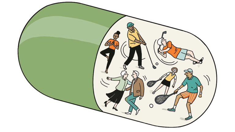
“Exercise is the magic pill,” says George Salem, noting that regular physical activity improves sleep and immunity, bolsters brain health and mental health, and decreases the chances of dying from any cause.

Over the past decade, Salem has conducted a series of studies on older adults, including veterans and prostate cancer survivors, who learn to play golf. He’s shown that in as few as 10 weeks, participation in the sport — which gets players striding up and down inclines and squatting to place and recover balls — increases walking ability and balance, improves performance on cognitive and memory tests, and reduces blood markers of inflammation.

The mental and social aspects of golf contribute to these benefits. As golfers strategize shots and fraternize with their peers, they’re stimulating their brains and engaging in what keeps Salem himself committed to the sport: play. “It’s more fun to play than it is to exercise,” he says.

Many of the participants in Salem’s studies express a desire to continue golfing after the introductory lessons end. “Once you’re hooked, you’re hooked,” Salem says. “Golfers will go out in the pouring rain and out in the snow. They show up because they’re playing with friends who expect them to show up.”

That level of investment in a fitness-boosting activity may help explain why golfers live five years longer on average than nongolfers. But if teeing off isn’t your cup of tea, don’t despair. “Other recreational activities, including pickleball and ballroom dancing, are likely just as beneficial,” Salem notes.

Illustration by Drue Wagner

Ready, Tech, Go!

NO MATTER HOW physically fit you are, sedentary behavior poses a significant threat to longevity. Sitting for long periods of time without breaks has been linked to a higher risk of heart disease, diabetes, cancer and death — even among those who exercise. Older adults with mobility issues, chronic pain and other health conditions are especially vulnerable to sitting for hours upon end.

At the Healthy Aging Research and Technology Lab in the USC Mrs. T.H. Chan Division of Occupational Science and Occupational Therapy, Stacey Schepens Niemiec, associate professor of research, is developing new tools to help older people reduce their amount of sedentary time. These tools incorporate both low-tech and high-tech solutions.

One is a smartphone app called Moving Up that tracks the number of hours users spend immobile and offers customized suggestions for integrating more movement into their self-reported activities. For example, if the user is watching television, the app may alert them to drink more water — not only for extra hydration, but to encourage more frequent walks to and from the bathroom.

Schepens Niemiec is also collaborating on the development of an active seating system that incorporates pedals — like a stationary bike, but without wheels — and sensors that track seated time. The system prompts users to pedal for a couple of minutes at regular intervals to counter the negative side effects of long periods of immobile sitting like stiffness and reduced blood flow.

It may seem against the grain to develop technological solutions for an older population, given that younger consumers are more often the assumed end users of the latest tech. But Schepens Niemiec is aiming to fill a gap. “The older adult population tends to get overlooked when it comes to tech-driven innovations,” she says. “Right from the start, I get older adults’ input in the design of the technology to help make it more accessible to them.”

Illustration of ears playing pickleball
Illustration by Drue Wagner

Now Hear This

ONE TECHNOLOGY for older adults that has greatly advanced in the past decade is hearing aids, which are smaller and have better sound quality than the devices of yesteryear. Yet only one in 10 of the approximately 40 million American adults who need hearing aids use them — likely due to a variety of factors, including cost and social stigma.

“A good number of patients tell me, ‘I don’t want to look old by wearing hearing aids,’” says Janet Choi, an otolaryngologyist with Keck Medicine and assistant professor at the Keck School of Medicine of USC.

That mindset can be self-defeating — because using hearing aids may help you live longer. Groundbreaking research by Choi and her collaborators revealed that among those with hearing loss, regular hearing aid users are almost 25% less likely to die from any cause than those who never wear them.

Though more research is needed to untangle why hearing aid use boosts lifespan, it’s possible that it confers protection against diseases linked to untreated hearing loss, including dementia and depression. “Some studies suggest that the sound input provided by hearing aids may have a positive impact on brain structures,” Choi says.

Choi is now researching ways to encourage hearing aid use among older people and counter social stigma associated with the devices, which she relates to from personal experience. The specialist in ear-related disorders was born with hearing loss in her left ear and now wears a hearing aid. But she didn’t do so as a child because her parents were worried she would be ridiculed at school.

It took Choi three tries before she found the right hearing aid for herself. Knowing that the time-intensive selection process can be a barrier to use, she’s developing an AI-driven database that helps match patients with hearing aids best suited to their unique needs.

Shore Up Support

Illustration by Drue Wagner

WHEN OLDER ADULTS with hearing loss adopt hearing aids, they’re better able to participate in conversations and stay engaged socially, buffering themselves from isolation. That itself may boost longevity: Feeling isolated makes it more likely you’ll develop dementia, depression, heart disease and stroke, and die prematurely.

Interdependence with others is critical throughout our lives. But as we age and need help with the tasks of daily living, interpersonal dynamics shift. Our adult children or spouse may step in to take care of us.

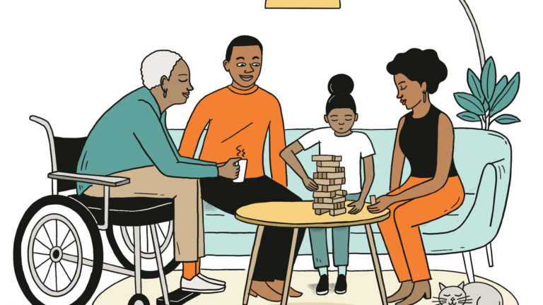
“That can be a wonderful new opportunity for a deepening love,” says Laura Mosqueda, professor of family medicine and geriatrics at Keck School of Medicine. “And it could also be an opportunity for abuse or neglect.”

One in 10 older adults is abused or neglected in their lifetime, and among those with Alzheimer’s or other dementias, the rate jumps to nearly one in two. Most commonly, the person carrying out the abuse is a close family member.

In her research on the causes of elder abuse, Mosqueda has found that abusive behavior is often rooted in caregivers’ difficulty coping with their demanding roles. “Eighty percent of abuse and neglect is preventable,” she says. “We need to look for the early warning signs: caregivers who are stressed, resentful, burdened and unhappy.”

Along with her collaborator Bonnie Olsen, professor of clinical family medicine and vice chair for research at Keck School of Medicine, Mosqueda is investigating how best to detect these signs and intervene by equipping caregivers with tactics for elder care and self-care. They’re one of several teams at USC working to support caregivers’ well-being and, by extension, the well-being of the older adults in their care.

At the USC Family Caregiver Support Center (FCSC) directed by Donna Benton, associate professor of gerontology, innovative research includes studies on how best to support dementia caregivers and how technology can improve caregiving. The FCSC also runs support groups and helplines for care-givers throughout the Los Angeles area, with groups such as the one at the First AME Church near the USC University Park Campus supporting diverse and low-income communities.

Tapping into the power of community to ease the challenges of modern aging is also a focus for María Aranda, professor in the USC Suzanne Dworak-Peck School of Social Work and director of the USC Edward R. Roybal Institute on Aging. Aranda conducts intervention research with English- and Spanish-speaking caregivers in the communities surrounding USC and has found that family caregivers of loved ones with medical conditions feel less alone in their journeys when they join supportive groups and share resources. Older adults who nurture community connections fare better, too, given the increased sense of purpose and emotional attachments.

“Whether it’s leisure activities, social activities related to their house of worship, or relationships with family, friends or neighbors, people who have a sense of purpose and meaning are likely to have a longer and more enjoyable life,” Aranda says.

Her insights affirm what Salem observed that pivotal day on the fairway. Social connectedness is a driving force of human thriving — whether on the golf course or over the course of a life well-lived.


Your Cells on Aging

Inside your body’s cells, structures called mitocho­ndria create the energy needed to carry out vital life processes. Cellular aging begins in these power­ houses. “Mitochondria are the first part of our cells to experience dysfunction as we age,” says Pinchas Cohen, dean of the USC Leonard Davis School of Ger­ontology and Distinguished Professor of gerontology, medicine and biological sciences.

Cohen and his collaborators have identified a series of microproteins produced by the mitochon­dria that naturally decrease with age and whose decline is linked to the onset of age­related diseases. His lab’s remarkable discoveries include mitochon­drial microproteins that protect against Alzheimer’s disease, Parkinson’s disease and diabetes, and are associated with longer life spans.

To sustain life, cells rely on a set of chemical reac­tions known as cell metabolism, which produces sub­stances called metabolites. Peter Mullen, assistant professor of molecular microbiology and immunology at the Keck School of Medicine and USC Leonard Davis, along with his collaborators, have identified multiple metabolites in the organs of mice that decrease sig­nificantly with age and whose reduction is associated with organ dysfunction. Many of these metabolites can also predict biological age in humans.

Both mitochondrial microproteins and metabo­lites have the potential to become tools of “precision longevity.” They may one day form the basis of drugs tailored to the specific cellular deficiencies of each individual to treat disease and prolong life. — Rachel B. Levin